
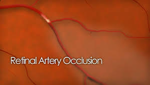
Retinal Artery Occlusions
What is a retinal artery occlusion?
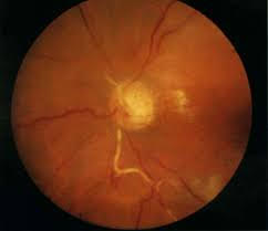
A retinal artery occlusion (RAO) occurs when an artery (blood vessel) is blocked. This stops the blood flow to the eye, which causes vision loss. The blockage is most commonly caused by atherosclerosis, or hardening of the blood vessels within the retina or it can travel from a blocked blood vessel elsewhere. The blockage can travel to the eye from the neck or the heart; or there can be clotting in the eye itself.
What are the symptoms of a retinal artery occlusion?
The most common symptom of an RAO is sudden, painless loss of vision in one eye, or in a portion of one eye. If the blockage is in the main central retinal artery, it will cause near-total vision loss. If the blockage is in one of the smaller arteries, it will cause partial vision loss, usually as a well-defined defect in the peripheral visual field. It can also block out either the top half or the bottom half of vision.
Who is at risk for a retinal artery occlusion?
Most patients who have an RAO are in their 60’s, however, it can occur in younger and older patients. Men are at a slightly higher risk. Patients with certain disorders such as atherosclerosis, heart disease, and some blood clotting disorders are also at a higher risk of having an RAO.
How is a retinal artery occlusion diagnosed?
An RAO is diagnosed primarily with a complete eye exam, including dilation of your pupils. Your ophthalmologist will carefully examine your retina. After diagnosis, you may need additional blood tests and/or imaging studies, such as an echocardiogram of the heart and a scan of the carotid arteries of the neck, in order to find the source of the blockage. Many times, once these causes are identified, they can be fixed in order to prevent further loss of vision or a stroke with more widespread effect/loss of function.
How is a retinal artery occlusion treated?
Once blockage of the artery occurs, there is limited time to restore any bloodflow. After approximately 90 minutes, the vision loss is permanent. Unfortunately, treatments are generally minimally effective. However, patients can try to massage the eye to dislodge or move the clot to smaller arteries resulting in less permanent damage to a person’s vision. The main treatment after an RAO is to determine the underlying cause and treat it to avoid further damage in the eye or elsewhere in the body.
What is the follow up after a retinal artery occlusion?
Patients should continue to see their primary care physicians on a regular basis to treat any risk factors and underlying disorders. It is also very important to continue follow up with an ophthalmologist because neovascularization, or formation of new vessels within the retina, can occur after an RAO. Neovascularization in the retinal or on the iris can lead to bleeding in the eye or very aggressive glaucoma. The ophthalmologist will monitor these patients with frequent, dilated eye exams.